Introduction
With the discovery of X-Rays by Physicist Wilhelm Conrad Roentgen in 1895, the first medical imaging technology became possible which is termed as Radiography. Wilhelm Conrad Roentgen also made the first X-ray images of human. Radiography defined the field of radiology.

Methodology
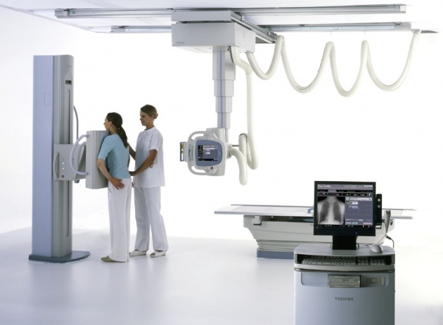
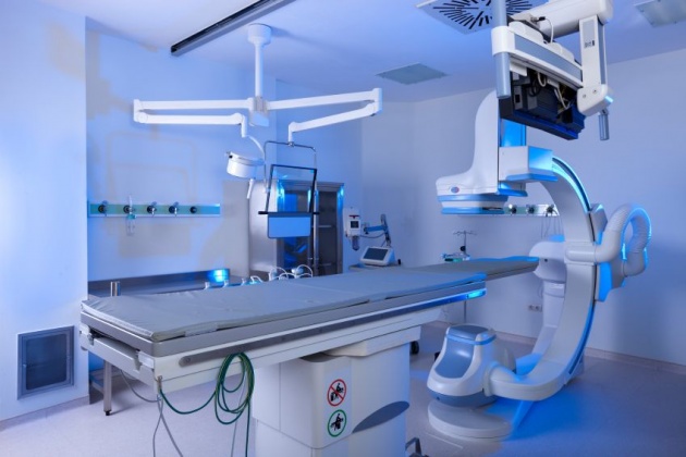
Radiography is performed with an X-ray source on one side of the patient and a typical flat X-ray detector on the other side (e.g film). A short duration (typically less than ½ second) pulse of X-rays is emitted by the X-ray source. Large fraction of the X-rays interacts with the patient while some rays pass through the patient and reach the detector, where the image forms.

Physics
The homogeneous distribution of x-rays which enters the patient is modified by scattering and absorption within the tissues and usually follows Lambert-Beer Law. The attenuation properties of tissues such as bone, soft tissues, and air inside the patient are very
different, resulting in a heterogeneous distribution of X-rays which emerges out of the patient. The X-ray image is that heterogeneous distribution of X-rays emerging out of the patient's body. The detector used in radiography can be
photographic plate (e.g. screen-film radiography) or an electronic detector system (i.e. digital radiography).

Applications
X-ray images are useful for a very wide range of medical indications, including the diagnosis of broken bones, lung cancer, cardiovascular disorders etc.
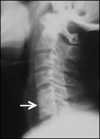
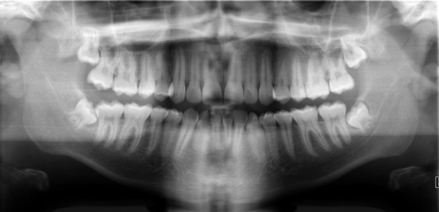
Some images